Saturday, December 22, 2007

Home Cinema Ready Made Solutions

01. ALL-IN-ONE, OR ‘ONE-BOX’ SYSTEMS

All-in-one home cinema systems are the simplest, most cost-effective way of reproducing the movie-going experience in the home. All-in-one systems include all you need to enjoy movies on DVD (apart from your display screen), just as the film director intended – with the same 5.1-channel surround sound as your local cinema.

Systems include DVD player and amplification, five ‘satellite’ speakers and a subwoofer speaker, for that essential blockbuster boom.

02. DVD/AMP + SPEAKERS

Many manufacturers are now selling DVD players with built-in amplification, or DVD/amps. This allows you to step up the home cinema ladder in stages, hooking the DVD/amp up to a TV before going full-on at a later date.

Full-on home cinema, of course, requires 5.1 speakers (five satellites and a subwoofer), and that’s all you need to complete the system.

03. DVD PLAYER + UPGRADE

If you’ve already got a DVD player, it’s easy to transform it into a complete home cinema system. Sure, you can go down the separates route, but there is an easier and cheaper way.

Some manufacturers are now producing complete DVD upgrade systems comprising the essential 5.1 speakers and amplification – just plug in your DVD player.


http://www.hdtvorg.co.uk/technology/home_cinema_ready_made.htm

HDTV Home Cinema

Home Cinema Introduction

The excitement of the cinema, from the comfort of your sofa... Home cinema brings the full sound and big-screen experience of the movie theatre into your living room. Technology has moved on so much in the last few years that you can enjoy amazing quality audiovisuals without paying a fortune. Dolby Digital Surround Sound allows you to immerse yourself in the action, while large plasma and LCD TVs offer incredibly clear widescreen viewing.

Almost any TV will work with a home theater system, but for the best possible experience, a larger screen is the way to go.

For optimal movie viewing, you'll need a "wide-screen" set with a 16:9 (width to height) aspect ratio. These most closely duplicate cinema screens, so you'll see the movie as the filmmakers intended. HDTV and most DVDs play in the wide-screen format.

When planning your home cinema system you can go for a ready made (out of the box) solution or you can build your own from seperate components. We will look at following these two different routes in the following sections. As sound plays such an important element of your home cinema system we also have a section dedicated to optimizing your systems sound.


http://www.hdtvorg.co.uk/technology/home_cinema.htm

HD Technology Accessories

1. Cables, dongles, and adapters

Of course, you will need a way to connect all of your high-def products. Some people argue that you should budget hundreds of pounds to pay for high-end premium cables in your home theater. We disagree. For 99 percent of the uses out there, inexpensive video cables perform just as well as high-end versions, for a fraction of the price.

The lone exception is for long cable runs - anything longer than 6 feet or 2 meters, where a name brand often offers greater dependability. No matter how much you spend, though, you'll likely want to have most of these on hand to make your HD connection odyssey as smooth as possible:

HDMI cable

HDMI cable (HDMI delivers audio and video over a single cable): If you have a state-of-the-art A/V system that uses HDMI jacks all around, you'll need to have plenty of cables on hand to connect each of your components (HDTV monitor, HD cable/satellite box, upscaling DVD player, and A/V receiver) to one another. HDMI cables are available in multiple lengths.

HDMI to DVI cable

HDMI-to-DVI cable (Perfect for connecting a DVI-equipped TV to an HDMI source) (HDMI delivers audio and video over a single cable) cable: If you have a state-of-the-art A/V system that uses HDMI jacks all around, you'll need to have plenty of cables on hand to connect each of your components (HDTV monitor, HD cable/satellite box, upscaling DVD player, and A/V receiver) to one another. HDMI cables are available in multiple lengths.

DVI to HDMI adapter

HDMI-to-DVI adapter (Turn any DVI jack into an HDMI connection): If you have a state-of-the-art A/V system that uses HDMI jacks all around, you'll need to have plenty of cables on hand to connect each of your components (HDTV monitor, HD cable/satellite box, upscaling DVD player, and A/V receiver) to one another. HDMI cables are available in multiple lengths.


2. HDMI switcher

HDMI switcher

Let's say you have multiple HDMI sources but only a single input on your HDTV. Or you have an older A/V receiver that lacks HDMI connections. Or you have an HDMI receiver, but you simply need more HDMI inputs. For each of these problems, an HDMI switcher is a suitable solution. A switcher (or just "switch") lets you toggle among as many as four HDMI inputs at the touch of a button.


http://www.hdtvorg.co.uk/technology/accessories.htm

CES 2008 Preview: HDTVs

There will be evolutionary improvements that focus on improving picture quality, as well as the emergence of one new display technology.

CES 2008 Coverage

The start of a new year is a signal for me to make my annual trek to the Consumer Electronics Show (CES) in Las Vegas where I'll inspect, first hand, the latest high-definition televisions. Every HDTV manufacturer I've spoken with recently has hinted that they'll be showcasing brand new models that will feature mostly evolutionary improvements. Their primary focus will be on improving picture quality. And at least one display technology should finally make the leap from the laboratory into the hands of awaiting consumers.

The liquid crystal display (LCD) dominates the flat-panel television category, and 2007 marked LCD's superseding the venerable cathode ray tube (CRT) as the most often-purchased type of TV. LCDs have long suffered from several image quality weaknesses that include mediocre contrast, color quality, and performance with fast moving imagery. The use of advanced backlight technologies, such as light emitting diodes (LEDs), in LCD panels can effectively address these problem areas while reducing the amount of mercury that is used in traditional fluorescent tube backlight systems. Dolby Labs' recent acquisition of a company that specialized in advanced LED backlight systems is the latest sign that the industry as a whole is seriously considering this technology as way to differentiate new products in an increasingly crowded field of brand names. Samsung and Sony have previously demonstrated large format liquid crystal HDTVs that feature LED backlight technology, and CES 2008 will certainly feature an expansion of these product lines. However, LED backlit LCDs will continue to command premium prices for the foreseeable future, and the show floor will certainly showcase new LCD designs that utilize improved fluorescent tube technology, as well as new screen and filter materials that offer further improvements in image quality at increasingly affordable prices.

The rear-projection television (RPTV) isn't dead yet, and this value-driven display category will receive some interesting updates at CES. Mitsubishi is expected to announce the availability of a laser illuminated DLP-based rear-projection television that the company first demonstrated at CES 2007. The original demo I previewed produced a picture that was too colorful (extreme color saturation), but if tamed, this laser-based display system has the potential to deliver exceptional color purity with excellent energy efficiency. I'll also be on the lookout for an update to Sony's prototype laser-lit RPTV using the company's SXRD microdisplay technology that also made an appearance at the 2007 show. The longevity of an RPTV's lamp module is a concern among many potential owners, and technologies, such as LED arrays and Luxim's LIFI hybrid light source that stimulates a sealed bulb with RF energy, are making lamp longevity a non-issue. This year's show will highlight more RPTVs that incorporate these light sources, and I'm curious to see if the latest LED arrays are able to match the brightness of traditional lamp-driven systems.

Now that every major plasma television manufacturer is producing panels with full 1080p resolution (1,920 by 1,080 pixels progressively scanned), I'm expecting the new models on display at CES to feature improved video processing with 24p video material (most film and digital cinema) as it is converted for display at the 60 Hz refresh rate that most TVs use—Pioneer is still the only TV manufacturer that offers a 72-Hz display mode for 3x frame processing of 24p material—eliminating a shaking artifact known as judder. Minimizing ambient light reflecting from the surface of a plasma television screen improves its appearance in rooms where lighting is uncontrollable—something most LCDs handle quite well. CES 2007 showcased the impressively non-reflective characteristics of new screen filters from Pioneer and Samsung. Pioneer's new KURO series of plasma TVs incorporates the company's latest filter technology, and I'm expecting some of the other primary plasma TV manufacturers including LG, Panasonic, and Samsung to demonstrate new models that can provide an alternative to LCDs for viewing in a well-lit environment.

Organic light emitting diode (OLED) displays should attract crowds of drooling onlookers once again. OLED displays can deliver an inky dark black for superb apparent contrast and color saturation, and they do so with a screen that measures only a few millimeters thick as it requires no backlight system like that used with LCD technology. Sony recently started offering an 11-inch OLED TV called the XEL-1 in Japan, and the company demonstrated a 27-inch 1080p resolution prototype at the 2007 show. Samsung is also keen on OLED development, and it seems likely that bigger and better OLED TVs are in the future for 2008.


http://www.pcmag.com/article2/0,2704,2239374,00.asp

Crunch-Time in High-Definition Format War

Performance of both Blu-ray and HD DVD during this second holiday season is crucial in establishing the new generation of optical discs.


Christmas 2007 could be the deciding joust for the fortunes of competing high-definition disc standards, Blu-Ray and HD DVD in the U.S., analysts at Understanding & Solutions claim.

Performance of both Blu-ray and HD DVD during this second holiday season is crucial in establishing the new generation of optical discs, and may bring the market closer to resolving the war between the competing formats.

"Blu-ray and HD DVD player prices have been falling since the summer, culminating in Toshiba's loss-leading sub-$100 HD DVD player, available in the USA last month for a limited time," said Jeremy Wills, consultant at Understanding & Solutions. "Price reductions in the US have continued into December, with Blu-ray players dropping below $300 for the first time, and HD DVD players below $200."

The analysts observe that drive, chipset and other system component prices are now falling as demand increases, speculating that manufacturers will be able to put together players for either format for under $150 at some point next year. This should translate into cheaper devices and wider adoption of them, with analysts expecting players to cost under $100 by 2011, should both formats remain,

"Blu-ray benefits from stronger Hollywood studio support and represents a greater proportion of High Definition disc production volumes and disc sales. To date, Paramount's move to sole support of HD DVD has failed to turn the market," Wills observes.

"As demand grows and manufacturing volumes build, we're going to see the costs of releasing on two different formats really start to bite. There may be surprises just around the corner, and we could see a lot more format clarity in 2008."

Despite all this action in the disc market, consumer confusion still persists. The importance of providing a coherent message through strong retailer support is essential, as many buyers still don't know what additional inputs are required in order to view High Definition content on a High Definition TV.


http://www.pcworld.com/article/id,140765-c,dvdtechnology/article.html

DNA - Interactions with proteins

All the functions of DNA depend on interactions with proteins. These protein interactions can be non-specific, or the protein can bind specifically to a single DNA sequence. Enzymes can also bind to DNA and of these, the polymerases that copy the DNA base sequence in transcription and DNA replication are particularly important.

DNA-binding proteins

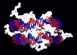
Interaction of DNA with histones (shown in white, top). These proteins' basic amino acids (below left, blue) bind to the acidic phosphate groups on DNA (below right, red).

Structural proteins that bind DNA are well-understood examples of non-specific DNA-protein interactions. Within chromosomes, DNA is held in complexes with structural proteins. These proteins organize the DNA into a compact structure called chromatin. In eukaryotes this structure involves DNA binding to a complex of small basic proteins called histones, while in prokaryotes multiple types of proteins are involved.[72][73] The histones form a disk-shaped complex called a nucleosome, which contains two complete turns of double-stranded DNA wrapped around its surface. These non-specific interactions are formed through basic residues in the histones making ionic bonds to the acidic sugar-phosphate backbone of the DNA, and are therefore largely independent of the base sequence.[74] Chemical modifications of these basic amino acid residues include methylation, phosphorylation and acetylation.[75] These chemical changes alter the strength of the interaction between the DNA and the histones, making the DNA more or less accessible to transcription factors and changing the rate of transcription.[76] Other non-specific DNA-binding proteins found in chromatin include the high-mobility group proteins, which bind preferentially to bent or distorted DNA.[77] These proteins are important in bending arrays of nucleosomes and arranging them into more complex chromatin structures.[78]

A distinct group of DNA-binding proteins are the single-stranded-DNA-binding proteins that specifically bind single-stranded DNA. In humans, replication protein A is the best-characterised member of this family and is essential for most processes where the double helix is separated, including DNA replication, recombination and DNA repair.[79] These binding proteins seem to stabilize single-stranded DNA and protect it from forming stem loops or being degraded by nucleases.

The lambda repressor helix-turn-helix transcription factor bound to its DNA target
The lambda repressor helix-turn-helix transcription factor bound to its DNA target[80]

In contrast, other proteins have evolved to specifically bind particular DNA sequences. The most intensively studied of these are the various classes of transcription factors, which are proteins that regulate transcription. Each one of these proteins bind to one particular set of DNA sequences and thereby activates or inhibits the transcription of genes with these sequences close to their promoters. The transcription factors do this in two ways. Firstly, they can bind the RNA polymerase responsible for transcription, either directly or through other mediator proteins; this locates the polymerase at the promoter and allows it to begin transcription.[81] Alternatively, transcription factors can bind enzymes that modify the histones at the promoter; this will change the accessibility of the DNA template to the polymerase.[82]

As these DNA targets can occur throughout an organism's genome, changes in the activity of one type of transcription factor can affect thousands of genes.[83] Consequently, these proteins are often the targets of the signal transduction processes that mediate responses to environmental changes or cellular differentiation and development. The specificity of these transcription factors' interactions with DNA come from the proteins making multiple contacts to the edges of the DNA bases, allowing them to "read" the DNA sequence. Most of these base-interactions are made in the major groove, where the bases are most accessible.[84]

The restriction enzyme EcoRV (green) in a complex with its substrate DNA
The restriction enzyme EcoRV (green) in a complex with its substrate DNA[85]

DNA-modifying enzymes

Nucleases and ligases

Nucleases are enzymes that cut DNA strands by catalyzing the hydrolysis of the phosphodiester bonds. Nucleases that hydrolyse nucleotides from the ends of DNA strands are called exonucleases, while endonucleases cut within strands. The most frequently-used nucleases in molecular biology are the restriction endonucleases, which cut DNA at specific sequences. For instance, the EcoRV enzyme shown to the left recognizes the 6-base sequence 5′-GAT|ATC-3′ and makes a cut at the vertical line. In nature, these enzymes protect bacteria against phage infection by digesting the phage DNA when it enters the bacterial cell, acting as part of the restriction modification system.[86] In technology, these sequence-specific nucleases are used in molecular cloning and DNA fingerprinting.

Enzymes called DNA ligases can rejoin cut or broken DNA strands, using the energy from either adenosine triphosphate or nicotinamide adenine dinucleotide.[87] Ligases are particularly important in lagging strand DNA replication, as they join together the short segments of DNA produced at the replication fork into a complete copy of the DNA template. They are also used in DNA repair and genetic recombination.[87]

Topoisomerases and helicases

Topoisomerases are enzymes with both nuclease and ligase activity. These proteins change the amount of supercoiling in DNA. Some of these enzyme work by cutting the DNA helix and allowing one section to rotate, thereby reducing its level of supercoiling; the enzyme then seals the DNA break.[27] Other types of these enzymes are capable of cutting one DNA helix and then passing a second strand of DNA through this break, before rejoining the helix.[88] Topoisomerases are required for many processes involving DNA, such as DNA replication and transcription.[28]

Helicases are proteins that are a type of molecular motor. They use the chemical energy in nucleoside triphosphates, predominantly ATP, to break hydrogen bonds between bases and unwind the DNA double helix into single strands.[89] These enzymes are essential for most processes where enzymes need to access the DNA bases.

Polymerases

Polymerases are enzymes that synthesise polynucleotide chains from nucleoside triphosphates. They function by adding nucleotides onto the 3′ hydroxyl group of the previous nucleotide in the DNA strand. As a consequence, all polymerases work in a 5′ to 3′ direction.[90] In the active site of these enzymes, the nucleoside triphosphate substrate base-pairs to a single-stranded polynucleotide template: this allows polymerases to accurately synthesise the complementary strand of this template. Polymerases are classified according to the type of template that they use.

In DNA replication, a DNA-dependent DNA polymerase makes a DNA copy of a DNA sequence. Accuracy is vital in this process, so many of these polymerases have a proofreading activity. Here, the polymerase recognizes the occasional mistakes in the synthesis reaction by the lack of base pairing between the mismatched nucleotides. If a mismatch is detected, a 3′ to 5′ exonuclease activity is activated and the incorrect base removed.[91] In most organisms DNA polymerases function in a large complex called the replisome that contains multiple accessory subunits, such as the DNA clamp or helicases.[92]

RNA-dependent DNA polymerases are a specialised class of polymerases that copy the sequence of an RNA strand into DNA. They include reverse transcriptase, which is a viral enzyme involved in the infection of cells by retroviruses, and telomerase, which is required for the replication of telomeres.[93][41] Telomerase is an unusual polymerase because it contains its own RNA template as part of its structure.[42]

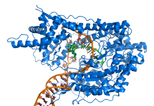
Transcription is carried out by a DNA-dependent RNA polymerase that copies the sequence of a DNA strand into RNA. To begin transcribing a gene, the RNA polymerase binds to a sequence of DNA called a promoter and separates the DNA strands. It then copies the gene sequence into a messenger RNA transcript until it reaches a region of DNA called the terminator, where it halts and detaches from the DNA. As with human DNA-dependent DNA polymerases, RNA polymerase II, the enzyme that transcribes most of the genes in the human genome, operates as part of a large protein complex with multiple regulatory and accessory subunits.[94]


http://en.wikipedia.org/wiki/DNA

DNA - Overview of biological functions

DNA usually occurs as linear chromosomes in eukaryotes, and circular chromosomes in prokaryotes. The set of chromosomes in a cell makes up its genome; the human genome has approximately 3 billion base pairs of DNA arranged into 46 chromosomes.[62] The information carried by DNA is held in the sequence of pieces of DNA called genes. Transmission of genetic information in genes is achieved via complementary base pairing. For example, in transcription, when a cell uses the information in a gene, the DNA sequence is copied into a complementary RNA sequence through the attraction between the DNA and the correct RNA nucleotides. Usually, this RNA copy is then used to make a matching protein sequence in a process called translation which depends on the same interaction between RNA nucleotides. Alternatively, a cell may simply copy its genetic information in a process called DNA replication. The details of these functions are covered in other articles; here we focus on the interactions between DNA and other molecules that mediate the function of the genome.

Genome structure

Genomic DNA is located in the cell nucleus of eukaryotes, as well as small amounts in mitochondria and chloroplasts. In prokaryotes, the DNA is held within an irregularly shaped body in the cytoplasm called the nucleoid.[63] The genetic information in a genome is held within genes. A gene is a unit of heredity and is a region of DNA that influences a particular characteristic in an organism. Genes contain an open reading frame that can be transcribed, as well as regulatory sequences such as promoters and enhancers, which control the expression of the open reading frame.

In many species, only a small fraction of the total sequence of the genome encodes protein. For example, only about 1.5% of the human genome consists of protein-coding exons, with over 50% of human DNA consisting of non-coding repetitive sequences.[64] The reasons for the presence of so much non-coding DNA in eukaryotic genomes and the extraordinary differences in genome size, or C-value, among species represent a long-standing puzzle known as the "C-value enigma."[65] However, DNA sequences that do not code protein may still encode functional non-coding RNA molecules, which are involved in the regulation of gene expression.[66]

T7 RNA polymerase (blue) producing a mRNA (green) from a DNA template (orange).
T7 RNA polymerase (blue) producing a mRNA (green) from a DNA template (orange).[67]

Some non-coding DNA sequences play structural roles in chromosomes. Telomeres and centromeres typically contain few genes, but are important for the function and stability of chromosomes.[42][68] An abundant form of non-coding DNA in humans are pseudogenes, which are copies of genes that have been disabled by mutation.[69] These sequences are usually just molecular fossils, although they can occasionally serve as raw genetic material for the creation of new genes through the process of gene duplication and divergence.[70]

Transcription and translation

A gene is a sequence of DNA that contains genetic information and can influence the phenotype of an organism. Within a gene, the sequence of bases along a DNA strand defines a messenger RNA sequence, which then defines one or more protein sequences. The relationship between the nucleotide sequences of genes and the amino-acid sequences of proteins is determined by the rules of translation, known collectively as the genetic code. The genetic code consists of three-letter 'words' called codons formed from a sequence of three nucleotides (e.g. ACT, CAG, TTT).

In transcription, the codons of a gene are copied into messenger RNA by RNA polymerase. This RNA copy is then decoded by a ribosome that reads the RNA sequence by base-pairing the messenger RNA to transfer RNA, which carries amino acids. Since there are 4 bases in 3-letter combinations, there are 64 possible codons (43 combinations). These encode the twenty standard amino acids, giving most amino acids more than one possible codon. There are also three 'stop' or 'nonsense' codons signifying the end of the coding region; these are the TAA, TGA and TAG codons.

DNA replication. The double helix is unwound by a helicase and topoisomerase. Next, one DNA polymerase produces the leading strand copy. Another DNA polymerase binds to the lagging strand. This enzyme makes discontinuous segments (called Okazaki fragments) before DNA ligase joins them together.

Replication

Cell division is essential for an organism to grow, but when a cell divides it must replicate the DNA in its genome so that the two daughter cells have the same genetic information as their parent. The double-stranded structure of DNA provides a simple mechanism for DNA replication. Here, the two strands are separated and then each strand's complementary DNA sequence is recreated by an enzyme called DNA polymerase. This enzyme makes the complementary strand by finding the correct base through complementary base pairing, and bonding it onto the original strand. As DNA polymerases can only extend a DNA strand in a 5′ to 3′ direction, different mechanisms are used to copy the antiparallel strands of the double helix.[71] In this way, the base on the old strand dictates which base appears on the new strand, and the cell ends up with a perfect copy of its DNA.


http://en.wikipedia.org/wiki/DNA

DNA - Chemical modifications

cytosine 5-methylcytosine thymine
Structure of cytosine with and without the 5-methyl group. After deamination the 5-methylcytosine has the same structure as thymine

Base modifications

The expression of genes is influenced by the chromatin structure of a chromosome and regions of heterochromatin (low or no gene expression) correlate with the methylation of cytosine. For example, cytosine methylation, to produce 5-methylcytosine, is important for X-chromosome inactivation.[47] The average level of methylation varies between organisms, with Caenorhabditis elegans lacking cytosine methylation, while vertebrates show higher levels, with up to 1% of their DNA containing 5-methylcytosine.[48] Despite the biological role of 5-methylcytosine it is susceptible to spontaneous deamination to leave the thymine base, and methylated cytosines are therefore mutation hotspots.[49] Other base modifications include adenine methylation in bacteria and the glycosylation of uracil to produce the "J-base" in kinetoplastids.[50][51]

DNA damage

Benzopyrene, the major mutagen in tobacco smoke, in an adduct to DNA.
Benzopyrene, the major mutagen in tobacco smoke, in an adduct to DNA.[52]

DNA can be damaged by many different sorts of mutagens. These include oxidizing agents, alkylating agents and also high-energy electromagnetic radiation such as ultraviolet light and x-rays. The type of DNA damage produced depends on the type of mutagen. For example, UV light mostly damages DNA by producing thymine dimers, which are cross-links between adjacent pyrimidine bases in a DNA strand.[53] On the other hand, oxidants such as free radicals or hydrogen peroxide produce multiple forms of damage, including base modifications, particularly of guanosine, as well as double-strand breaks.[54] It has been estimated that in each human cell, about 500 bases suffer oxidative damage per day.[55][56] Of these oxidative lesions, the most dangerous are double-strand breaks, as these lesions are difficult to repair and can produce point mutations, insertions and deletions from the DNA sequence, as well as chromosomal translocations.[57]

Many mutagens intercalate into the space between two adjacent base pairs. Intercalators are mostly aromatic and planar molecules, and include ethidium, daunomycin, doxorubicin and thalidomide. In order for an intercalator to fit between base pairs, the bases must separate, distorting the DNA strands by unwinding of the double helix. These structural changes inhibit both transcription and DNA replication, causing toxicity and mutations. As a result, DNA intercalators are often carcinogens, with benzopyrene diol epoxide, acridines, aflatoxin and ethidium bromide being well-known examples.[58][59][60] Nevertheless, due to their properties of inhibiting DNA transcription and replication, they are also used in chemotherapy to inhibit rapidly-growing cancer cells.[61]


http://en.wikipedia.org/wiki/DNA

DNA - Physical and chemical properties

The chemical structure of DNA.

DNA is a long polymer made from repeating units called nucleotides.[1][2] The DNA chain is 22 to 26 Ångströms wide (2.2 to 2.6 nanometres), and one nucleotide unit is 3.3 Ångstroms (0.33 nanometres) long.[3] Although each individual repeating unit is very small, DNA polymers can be enormous molecules containing millions of nucleotides. For instance, the largest human chromosome, chromosome number 1, is 220 million base pairs long.[4]

In living organisms, DNA does not usually exist as a single molecule, but instead as a tightly-associated pair of molecules.[5][6] These two long strands entwine like vines, in the shape of a double helix. The nucleotide repeats contain both the segment of the backbone of the molecule, which holds the chain together, and a base, which interacts with the other DNA strand in the helix. In general, a base linked to a sugar is called a nucleoside and a base linked to a sugar and one or more phosphate groups is called a nucleotide. If multiple nucleotides are linked together, as in DNA, this polymer is referred to as a polynucleotide.[7]

The backbone of the DNA strand is made from alternating phosphate and sugar residues.[8] The sugar in DNA is 2-deoxyribose, which is a pentose (five-carbon) sugar. The sugars are joined together by phosphate groups that form phosphodiester bonds between the third and fifth carbon atoms of adjacent sugar rings. These asymmetric bonds mean a strand of DNA has a direction. In a double helix the direction of the nucleotides in one strand is opposite to their direction in the other strand. This arrangement of DNA strands is called antiparallel. The asymmetric ends of DNA strands are referred to as the 5′ (five prime) and 3′ (three prime) ends. One of the major differences between DNA and RNA is the sugar, with 2-deoxyribose being replaced by the alternative pentose sugar ribose in RNA.[6]

The DNA double helix is stabilized by hydrogen bonds between the bases attached to the two strands. The four bases found in DNA are adenine (abbreviated A), cytosine (C), guanine (G) and thymine (T). These four bases are shown below and are attached to the sugar/phosphate to form the complete nucleotide, as shown for adenosine monophosphate.

These bases are classified into two types; adenine and guanine are fused five- and six-membered heterocyclic compounds called purines, while cytosine and thymine are six-membered rings called pyrimidines.[6] A fifth pyrimidine base, called uracil (U), usually takes the place of thymine in RNA and differs from thymine by lacking a methyl group on its ring. Uracil is not usually found in DNA, occurring only as a breakdown product of cytosine, but a very rare exception to this rule is a bacterial virus called PBS1 that contains uracil in its DNA.[9] In contrast, following synthesis of certain RNA molecules, a significant number of the uracils are converted to thymines by the enzymatic addition of the missing methyl group. This occurs mostly on structural and enzymatic RNAs like transfer RNAs and ribosomal RNA.[10]

Major and minor grooves

Animation of the structure of a section of DNA. The bases lie horizontally between the two spiraling strands. Large version
Animation of the structure of a section of DNA. The bases lie horizontally between the two spiraling strands. Large version[11]

The double helix is a right-handed spiral. As the DNA strands wind around each other, they leave gaps between each set of phosphate backbones, revealing the sides of the bases inside (see animation). There are two of these grooves twisting around the surface of the double helix: one groove, the major groove, is 22 Å wide and the other, the minor groove, is 12 Å wide.[12] The narrowness of the minor groove means that the edges of the bases are more accessible in the major groove. As a result, proteins like transcription factors that can bind to specific sequences in double-stranded DNA usually make contacts to the sides of the bases exposed in the major groove.[13]

Base pairing

At top, a GC base pair with three hydrogen bonds. At the bottom, AT base pair with two hydrogen bonds. Hydrogen bonds are shown as dashed lines.

Each type of base on one strand forms a bond with just one type of base on the other strand. This is called complementary base pairing. Here, purines form hydrogen bonds to pyrimidines, with A bonding only to T, and C bonding only to G. This arrangement of two nucleotides binding together across the double helix is called a base pair. In a double helix, the two strands are also held together via forces generated by the hydrophobic effect and pi stacking, which are not influenced by the sequence of the DNA.[14] As hydrogen bonds are not covalent, they can be broken and rejoined relatively easily. The two strands of DNA in a double helix can therefore be pulled apart like a zipper, either by a mechanical force or high temperature.[15] As a result of this complementarity, all the information in the double-stranded sequence of a DNA helix is duplicated on each strand, which is vital in DNA replication. Indeed, this reversible and specific interaction between complementary base pairs is critical for all the functions of DNA in living organisms.[1]

The two types of base pairs form different numbers of hydrogen bonds, AT forming two hydrogen bonds, and GC forming three hydrogen bonds (see figures, left). The GC base pair is therefore stronger than the AT base pair. As a result, it is both the percentage of GC base pairs and the overall length of a DNA double helix that determine the strength of the association between the two strands of DNA. Long DNA helices with a high GC content have stronger-interacting strands, while short helices with high AT content have weaker-interacting strands.[16] Parts of the DNA double helix that need to separate easily, such as the TATAAT Pribnow box in bacterial promoters, tend to have sequences with a high AT content, making the strands easier to pull apart.[17] In the laboratory, the strength of this interaction can be measured by finding the temperature required to break the hydrogen bonds, their melting temperature (also called Tm value). When all the base pairs in a DNA double helix melt, the strands separate and exist in solution as two entirely independent molecules. These single-stranded DNA molecules have no single common shape, but some conformations are more stable than others.[18]

Sense and antisense

A DNA sequence is called "sense" if its sequence is the same as that of a messenger RNA copy that is translated into protein. The sequence on the opposite strand is complementary to the sense sequence and is therefore called the "antisense" sequence. Since RNA polymerases work by making a complementary copy of their templates, it is this antisense strand that is the template for producing the sense messenger RNA. Both sense and antisense sequences can exist on different parts of the same strand of DNA (i.e. both strands contain both sense and antisense sequences). In both prokaryotes and eukaryotes, antisense RNA sequences are produced, but the functions of these RNAs are not entirely clear.[19] One proposal is that antisense RNAs are involved in regulating gene expression through RNA-RNA base pairing.[20]

A few DNA sequences in prokaryotes and eukaryotes, and more in plasmids and viruses, blur the distinction made above between sense and antisense strands by having overlapping genes.[21] In these cases, some DNA sequences do double duty, encoding one protein when read 5′ to 3′ along one strand, and a second protein when read in the opposite direction (still 5′ to 3′) along the other strand. In bacteria, this overlap may be involved in the regulation of gene transcription,[22] while in viruses, overlapping genes increase the amount of information that can be encoded within the small viral genome.[23] Another way of reducing genome size is seen in some viruses that contain linear or circular single-stranded DNA as their genetic material.[24][25]

Supercoiling

DNA can be twisted like a rope in a process called DNA supercoiling. With DNA in its "relaxed" state, a strand usually circles the axis of the double helix once every 10.4 base pairs, but if the DNA is twisted the strands become more tightly or more loosely wound.[26] If the DNA is twisted in the direction of the helix, this is positive supercoiling, and the bases are held more tightly together. If they are twisted in the opposite direction, this is negative supercoiling, and the bases come apart more easily. In nature, most DNA has slight negative supercoiling that is introduced by enzymes called topoisomerases.[27] These enzymes are also needed to relieve the twisting stresses introduced into DNA strands during processes such as transcription and DNA replication.[28]

From left to right, the structures of A, B and Z DNA

Alternative double-helical structures

DNA exists in several possible conformations. The conformations so far identified are: A-DNA, B-DNA, C-DNA, D-DNA,[29] E-DNA,[30] H-DNA,[31] L-DNA,[29] P-DNA,[32] and Z-DNA.[8][33] However, only A-DNA, B-DNA, and Z-DNA have been observed in naturally occurring biological systems. Which conformation DNA adopts depends on the sequence of the DNA, the amount and direction of supercoiling, chemical modifications of the bases and also solution conditions, such as the concentration of metal ions and polyamines.[34] Of these three conformations, the "B" form described above is most common under the conditions found in cells.[35] The two alternative double-helical forms of DNA differ in their geometry and dimensions.

The A form is a wider right-handed spiral, with a shallow, wide minor groove and a narrower, deeper major groove. The A form occurs under non-physiological conditions in dehydrated samples of DNA, while in the cell it may be produced in hybrid pairings of DNA and RNA strands, as well as in enzyme-DNA complexes.[36][37] Segments of DNA where the bases have been chemically-modified by methylation may undergo a larger change in conformation and adopt the Z form. Here, the strands turn about the helical axis in a left-handed spiral, the opposite of the more common B form.[38] These unusual structures can be recognized by specific Z-DNA binding proteins and may be involved in the regulation of transcription.[39]

Structure of a DNA quadruplex formed by telomere repeats. The conformation of the DNA backbone diverges significantly from the typical helical structure
Structure of a DNA quadruplex formed by telomere repeats. The conformation of the DNA backbone diverges significantly from the typical helical structure[40]

Quadruplex structures

At the ends of the linear chromosomes are specialized regions of DNA called telomeres. The main function of these regions is to allow the cell to replicate chromosome ends using the enzyme telomerase, as the enzymes that normally replicate DNA cannot copy the extreme 3′ ends of chromosomes.[41] As a result, if a chromosome lacked telomeres it would become shorter each time it was replicated. These specialized chromosome caps also help protect the DNA ends from exonucleases and stop the DNA repair systems in the cell from treating them as damage to be corrected.[42] In human cells, telomeres are usually lengths of single-stranded DNA containing several thousand repeats of a simple TTAGGG sequence.[43]

These guanine-rich sequences may stabilize chromosome ends by forming very unusual structures of stacked sets of four-base units, rather than the usual base pairs found in other DNA molecules. Here, four guanine bases form a flat plate and these flat four-base units then stack on top of each other, to form a stable G-quadruplex structure.[44] These structures are stabilized by hydrogen bonding between the edges of the bases and chelation of a metal ion in the centre of each four-base unit. The structure shown to the left is a top view of the quadruplex formed by a DNA sequence found in human telomere repeats. The single DNA strand forms a loop, with the sets of four bases stacking in a central quadruplex three plates deep. In the space at the centre of the stacked bases are three chelated potassium ions.[45] Other structures can also be formed, with the central set of four bases coming from either a single strand folded around the bases, or several different parallel strands, each contributing one base to the central structure.

In addition to these stacked structures, telomeres also form large loop structures called telomere loops, or T-loops. Here, the single-stranded DNA curls around in a long circle stabilized by telomere-binding proteins.[46] At the very end of the T-loop, the single-stranded telomere DNA is held onto a region of double-stranded DNA by the telomere strand disrupting the double-helical DNA and base pairing to one of the two strands. This triple-stranded structure is called a displacement loop or D-loop.[44]


http://en.wikipedia.org/wiki/DNA

Deoxyribonucleic acid (DNA)

The structure of part of a DNA double helix

Deoxyribonucleic acid (DNA) is a nucleic acid that contains the genetic instructions used in the development and functioning of all known living organisms. The main role of DNA molecules is the long-term storage of information. DNA is often compared to a set of blueprints, since it contains the instructions needed to construct other components of cells, such as proteins and RNA molecules. The DNA segments that carry this genetic information are called genes, but other DNA sequences have structural purposes, or are involved in regulating the use of this genetic information.

Chemically, DNA is a long polymer of simple units called nucleotides, with a backbone made of sugars and phosphate groups joined by ester bonds. Attached to each sugar is one of four types of molecules called bases. It is the sequence of these four bases along the backbone that encodes information. This information is read using the genetic code, which specifies the sequence of the amino acids within proteins. The code is read by copying stretches of DNA into the related nucleic acid RNA, in a process called transcription. Most of these RNA molecules are used to synthesize proteins, but others are used directly in structures such as ribosomes and spliceosomes.

Within cells, DNA is organized into structures called chromosomes. These chromosomes are duplicated before cells divide, in a process called DNA replication. Eukaryotic organisms such as animals, plants, and fungi store their DNA inside the cell nucleus, while in prokaryotes such as bacteria it is found in the cell's cytoplasm. Within the chromosomes, chromatin proteins such as histones compact and organize DNA, which helps control its interactions with other proteins and thereby control which genes are transcribed.


http://en.wikipedia.org/wiki/DNA